Teeth are not only essential for chewing but they play an important role in speech and facial esthetics. They are also the hardest substances in the human body.
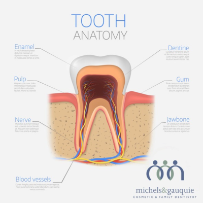 A tooth morphologically consists of several parts. The crown is the visible part of the tooth that projects into the oral cavity. The root that descends below the gum line, into the jaw. The root functions as an anchor for the tooth and has an intricate passage for blood and nerve supply that maintains the tooth viability. The crown is the surface that breaks down the food when opposing teeth are brought together on chewing. Both the crown and root consists of hard and soft tissue.
The hard tissue covering the crown is called enamel whereas the root is covered by cementum. Enamel is the hardest, white outer layer of the tooth, and is majorly composed of calcium phosphate. Cementum is softer compared to enamel. Cementum is a layer of hard connective tissue anchoring the roots of the teeth firmly to the gums and jawbone.
The next layer under both enamel and cementum is Dentin, making up the main bulk of the tooth. Dentin is a hard tissue, it is however much more porous than other hard tissues. This porous structure of dentin allows nutrients to be transferred through the tooth layers. Dentin contains microscopic tubules and if the enamel gets damaged, these microscopic tubes can conduct heat or cold and cause sensitivity or pain.
The pulp cavity housing the pulp tissue underlies the dentin. It has a rich blood supply and nerve supply, which is essential for maintaining tooth vitality. The tooth root lies embedded in bone, covered by the gingival tissue. The root is held in place by the tissue structure called periodontal ligaments that originates from the surrounding bone and is embedded into cementum.


